A) The Tissue System
We were discussing types of tissues based on the types of cells present. Let us now consider how tissues vary depending on their location in the plant body. Their structure and function would also be dependent on location. On the basis of their structure and location, there are three types of tissue systems. These are the epidermal tissue system, the ground or fundamental tissue system and the vascular or conducting tissue system.
i) Epidermal Tissue System
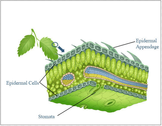
The epidermal tissue system forms the outer-most covering of the whole plant body and comprises epidermal cells, stomata and the epidermal appendages – the trichomes and hairs. The epidermis is the outermost layer of the primary plant body. It is made up of elongated, compact.
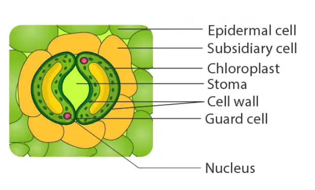
arranged cells, which form a continuous layer. Epidermis is usually single layered. Epidermal cells are parenchymatous with a small amount of cytoplasm lining the cell wall and a large vacuole. The outside of the epidermis is often covered with a waxy thick layer called the cuticle which prevents the loss of water. Cuticle is absent in roots. Stomata are structures present in the epidermis of leaves. Stomata regulate the process of transpiration and gaseous exchange. Each stoma is composed of two bean shaped cells known as guard cells which enclose stomatal pore. In grasses,
the guard cells are dumb-bell shaped. The outer walls of guard cells (away from the stomatal pore) are thin and the inner walls (towards the stomatal pore) are highly thickened. The guard cells possess chloroplasts and regulate the opening and closing of stomata. Sometimes, a few epidermal cells, in the vicinity of the guard cells become specialized in their shape and size and are known as subsidiary cells. The stomatal aperture, guard cells and the surrounding subsidiary cells are together called stomatal apparatus.
ii) The Ground Tissue System
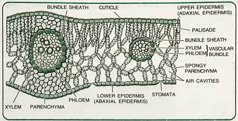
All tissues except epidermis and vascular bundles constitute the ground tissue. It consists of simple tissues such as parenchyma, collenchyma and sclerenchyma. Parenchymatous cells are usually present in cortex, pericycle, pith and medullary rays, in the primary stems and roots. In leaves, the ground tissue consists of thin-walled chloroplast containing cells and is called mesophyll.
iii) The Vascular Tissue System
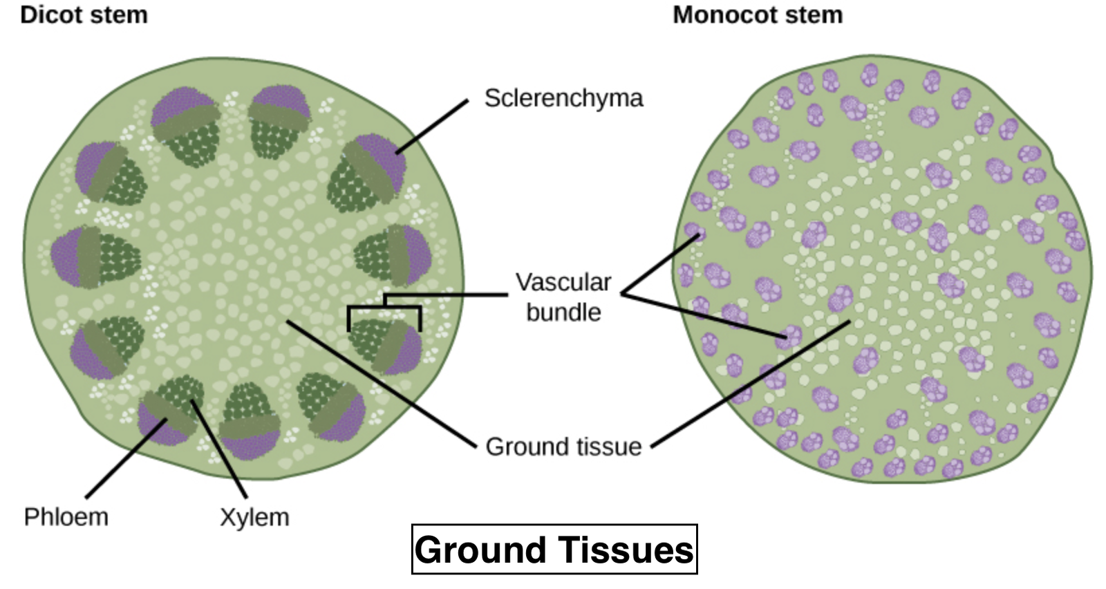
The vascular system consists of complex tissues, the phloem and the xylem. The xylem and phloem together constitute vascular bundles . In dicotyledonous stems, cambium is present between phloem and xylem. Such vascular bundles because of the presence of cambium possess the ability to form secondary xylem and phloem tissues, and hence are called open vascular bundles.
B. Anatomy of dicotyledonous and monocotyledonous plants
For a better understanding of tissue organization of roots, stems and leaves, it is convenient to study the transverse sections of the mature zones of these organs.
i) Dicotyledonous Root:
it shows the transverse section of the sunflower root. The internal tissue organisation is as follows: The outermost layer is epiblema. Many of the cells of epiblema protrude in the form of unicellular root hairs. The cortex consists of several layers of thin-walled parenchyma cells. with intercellular spaces. The innermost layer of the cortex is called endodermis. It comprises a single layer of barrelshaped cells without any intercellular spaces. The tangential as well as radial walls of the endodermal cells have a deposition of water-impermeable, waxy material suberin in the form of casparian strips.
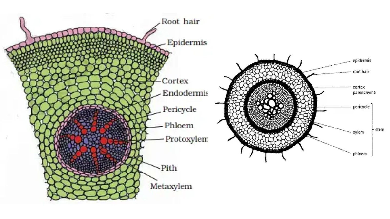
Next to endodermis lie a few layers of thick-walled parenchyomatous cells referred to as pericycle. Initiation of lateral roots and vascular cambium during the secondary growth takes place in these cells. The pith is small or inconspicuous. The parenchymatous cells which lie between the xylem and the phloem are called conjunctive tissue. There are usually two to four xylem and phloem patches. Later, a cambium ring develops between the xylem and phloem.
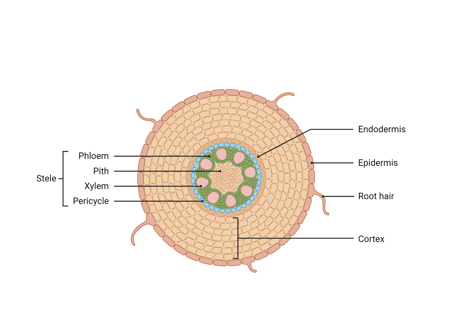
ii) Monocotyledonous Root
The anatomy of the monocot root is similar to the dicot root in many respects .Ithas epidermis, cortex, endodermis, pericycle, vascular bundles and pith. As compared to the dicot root which have fewer xylem bundles, there are usually more than six (polyarch) xylem bundles in the monocot root. Pith is large and well developed. Monocotyledonous roots do not undergo any secondary growth.
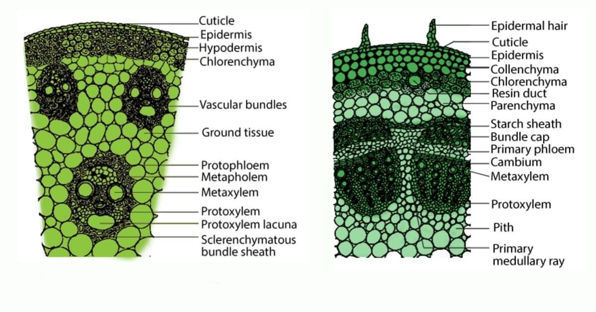
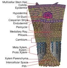
iii) Dicotyledonous Stem
The transverse section of a typical young dicotyledonous stem shows that the epidermis is the outermost protective layer of the stem. Covered with a thin layer of cuticle, it may bear trichomes and a few stomata. The cells arranged in multiple layers between epidermis and pericycle constitute the cortex. It consists of three sub-zones. The outer hypodermis, consists of a few layers of collenchymatous cells just below the epidermis, which provide mechanical strength to the young stem. Cortical layers below hypodermis consist of rounded thin walledparenchymatous cells with conspicuous intercellular spaces. The innermost layer of the cortex is called the endodermis. The cells of the endodermis are rich in starch grains and the layer is also referred to as the starch sheath.
Isobilateral (Monocotyledonous) Leaf
The anatomy of isobilateral leaf is similar to that of the dorsiventral leaf in many ways. It shows the following characteristic differences. In an isobilateral leaf, the stomata are present on both the surfaces of the epidermis; and the mesophyll is not differentiated into palisade and spongy parenchyma. In grasses, certain adaxial epidermal cells along the veins modify themselves
into large, empty, colourless cells. These are called bulliform cells. When the bulliform cells in the leaves have absorbed water and are turgid, the leaf surface is exposed. When they are flaccid due to water stress, they make the leaves curl inwards to minimize water loss. The parallel venation in monocot leaves is reflected in the near similar sizes of vascular bundles (except in main veins) as seen in vertical sections of the leaves.