There are two centrioles in every vertebrate cell. They support the cell while it divides. They function throughout the meiotic and mitotic processes. While they are absent from many fungi, angiosperms (flowering plants), and pinophyta (conifers), they may be present in some lower plants, such as Chlamydomonas. When the cell is not dividing, they are not visible; however, they are typically found close to the nucleus.
Centrioles outlook
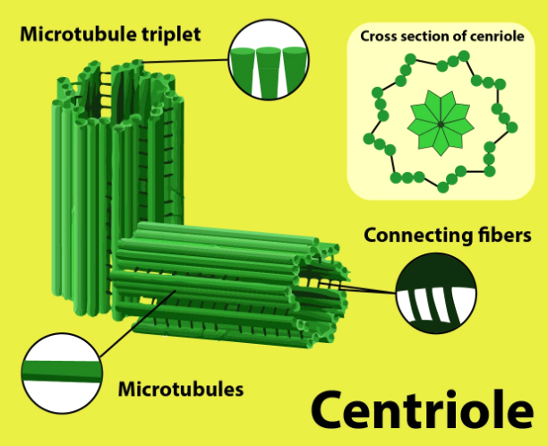
All the centrioles are formed of 9 groups of microtubule triplets organized in a cylindrical shape. The detailed structure of centrioles can be studied only under an electron microscope. These are associated together at right angles to each other.
- The embryo of Drosophila melanogasterand elegans are exceptions to this organization. The former forms 9 pairs instead of microtubule triplets, whereas the premature embryos and sperm cells of C. elegans have 9 single microtubules.
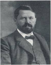
- Edouard van Beneden and Theodor Boveri observed and identified the centrioles for the first time in 1883 and 1888. The structure of the duplication of centrioles was first given by Joseph G. Gall and Etienne de Harven in the 1950s.
- The centriole helps in organizing the mitotic spindle and completes the process of cytokinesis. However, centrioles were believed to be necessary for the formation of the mitotic spindle in the animal cell.
- Although several recent types of research have explained that the cell that does not have a centriole (surgically removed through laser) can function without it in the G1 level of interphase, and can be formed later in a de novo manner.
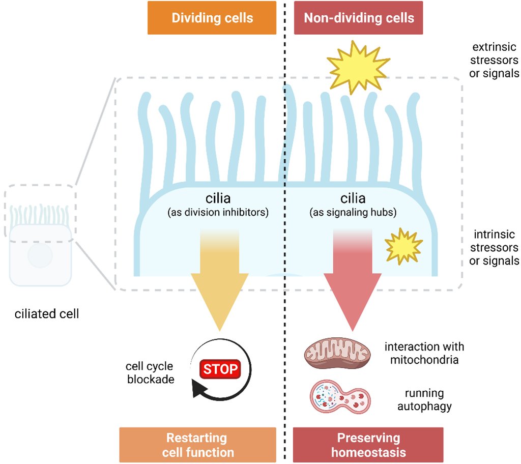
The location of the centrioles plays a key role in the three-dimensional organization of the cell, as it also regulates the location of the nucleus.
In flagellated and ciliated organisms, the location of such organelles is decided after the mother centrioles that form the base.
The Function of Centrioles
The important functions of centrioles are as follows:
1. The centrioles have the ability to create new centrioles despite lacking DNA.
2. They are capable of becoming basal bodies.
3. Cilia and flagella are produced by the basal bodies.
4. Through the formation of microtubule-organizing centers, they aid in cell division.
5. The distal centriole is the one that forms the axial filament or tail out of the two centrioles.
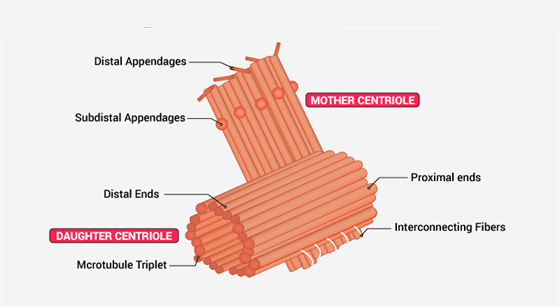
Centriole Structure
- Although some are as little as 0.16 μm and others as long as 8 μm, centrioles and basal bodies are cylindrical structures with a diameter of 0.15–0.25 μm and a length of typically 0.3–0.7 μm.
- Although they can be seen under a light microscope, only an electron microscope was able to expose the centriole’s structure.
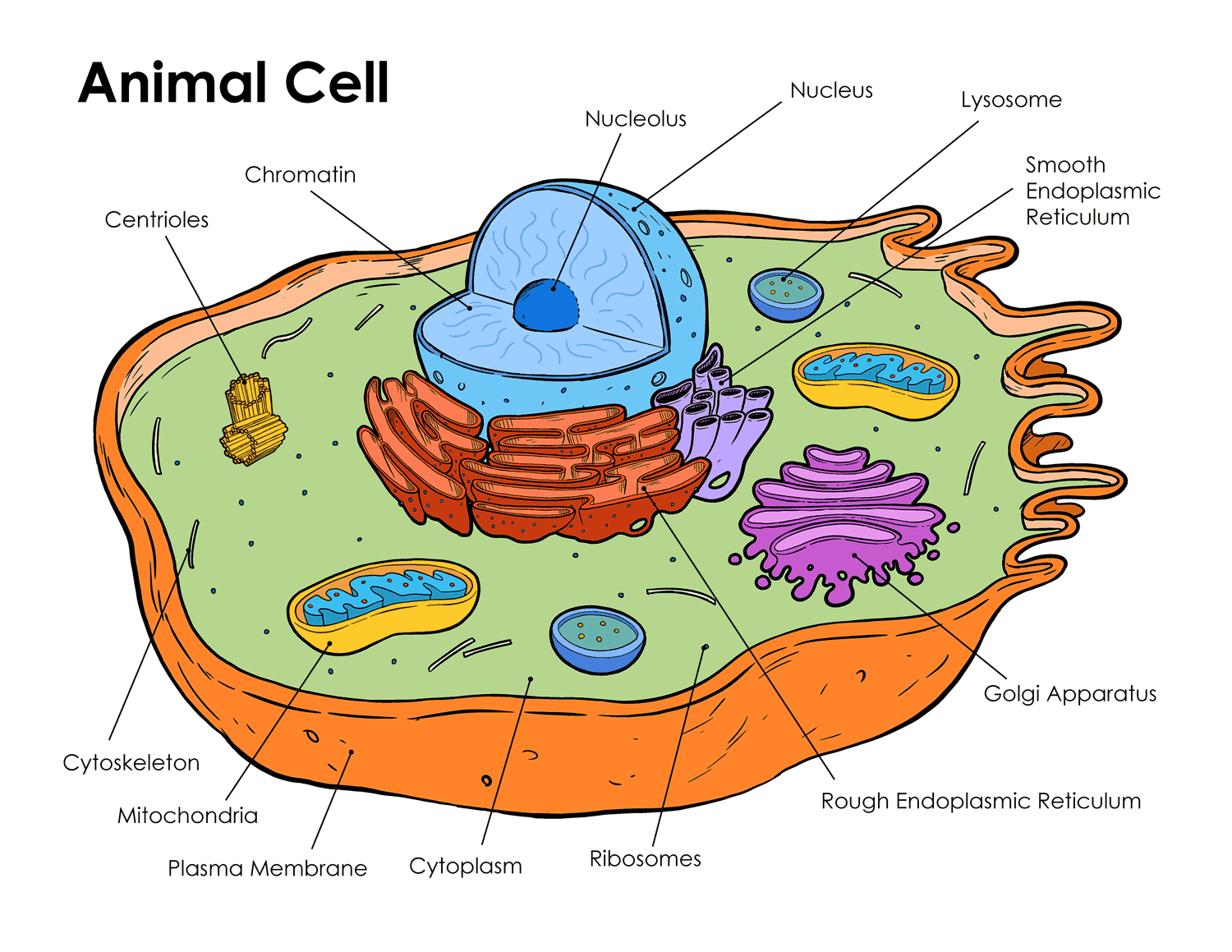
- The centrosome, which is located close to the nucleus, has two centrioles in every cell. One pair of centrioles has members that are perpendicular to one another.
Discovery of Centrioles
The presence of centrioles was observed in the late 19th century:
- Edouard van Beneden (1883) and Theodor Boveri (1888) first identified centrioles in animal cells.
- Later, in the 1950s, Joseph G. Gall and Etienne de Harven described how centrioles duplicate.
These discoveries were fundamental in understanding cell division mechanisms and the intricate organization of microtubules in eukaryotic cells.
Extra Thoughts and Particular Details
- Cell division is not immediately stopped when centrioles are removed experimentally (for instance, by laser ablation). Centrioles significantly improve spindle formation’s stability and efficiency, yet, since the process may become less ordered.
- The mother centriole directly produces the basal bodies that serve as the base of cilia or flagella in organisms with flagellation and ciliation.
Centrioles in Animal Cells
Centrioles are most commonly found in animal cells. They are crucial for forming spindle fibers during cell division (mitosis and meiosis). When an animal cell enters mitosis, each centriole pair migrates to opposite poles of the cell, contributing to the formation of spindle fibers that pull the chromosomes apart. This ensures the proper distribution of genetic material into the daughter cells.
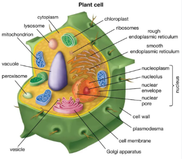
Centrioles in Plant Cells
Although it was once believed that centrioles were necessary for spindle formation, research shows that cells (under experimental conditions) can occasionally form spindles without centrioles. The majority of higher plant cells (angiosperms and conifers) lack centrioles, but some lower plant forms, such as Chlamydomonas, do have them. During mitosis in animal cells, the centriole duplicates in the S phase of the cell cycle, and spindle fibers start to form and attach to the chromosomes, separating them to ensure each daughter cell receives the correct set of genetic information.
In mitosis, centrioles
The centriole duplicates in the S phase of the cell cycle during animal cell mitosis. To guarantee that each daughter cell obtains the appropriate set of genetic information, spindle fibers start to develop and adhere to the chromosomes, dividing them. Research shows that cells can occasionally create spindles without centrioles, despite the long-held belief that centrioles are necessary for spindle production.