A cell envelope: what is it?
The cell membrane, cell wall, and outer membrane, if any, combine to form the cell envelope. This envelope is typically seen in prokaryotes, such as bacteria. It includes a bacterium’s inner and outer cell walls.
The cell’s structural integrity is provided by the cell envelope. It defends the cell in prokaryotes against internal turbo pressure brought on by a large concentration of macromolecules within the cell.
Bacterial cell envelopes come in two varieties:
- Gram-positive cell wall
- Gram-negative cell wall
Functions of Cell Envelope
Following are the important functions of the cell envelope:
- The bacterial cell wallnot only maintains the cell shape and prevents it from bursting or collapsing but, as observed in the case of motile bacteria, it forms filamentous extensions called flagella.
- The cell membrane also regulates the transport of substances in and out of the cell.
- A special structure known as embosomed is formed by an extension of the plasma membrane into the cell wall. These extensions are usually in the form of vesicles, tubules, and lamellae.
They help the cell with various functions like a synthesis of a cell wall, DNA replication, distribution of daughter cells, respiration, secretions, etc.
Capsule
The glycocalyx is thick and tough here. It is made up of polysaccharides and proteins. It protects the cell against desiccation, antibiotics and viral attack. Gelatinous glycocalyx glues together the bacterial cells to form colonies. It provides virulence also.
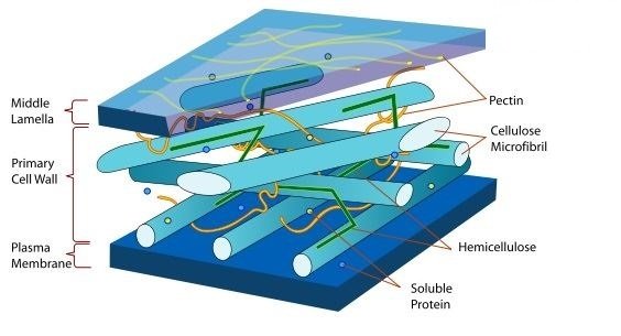
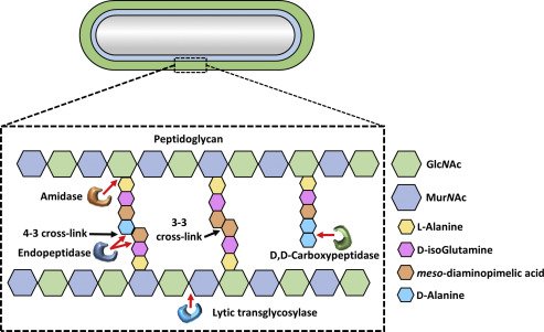
Cell wall
It is the middle layer of the cell envelope of a prokaryote which is sandwiched between the glycocalyx and cell membrane. Cell wall in bacteria is made up of peptidoglycan which contains polymers of modified sugars (such as N-acetyl glucosamine and N-acetyl muriatic acid) cross linked with some short peptides. Teichoic acid is also a component of the cell wall in some bacteria. Mycoplasma lacks a cell wall. Cell wall composition varies in gram negative and gram positive bacteria. Cell wall maintains shape and provides support and strength to the majority of the prokaryotic cells such as bacteria. It prevents the bacteria from bursting.
Membrane of a cell
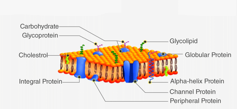
The innermost layer of the cell envelope is called the cell membrane. The membranes have a rather fluid quality. Certain molecules on each side of it can pass through it selectively. Sterols like cholesterol are absent from the majority of bacterial cell membranes. Hopanoids are pentacyclic compounds that can be found in the cell membranes of certain bacteria. They aid in stabilizing membranes’ molecular structure. It is composed of lipoprotein and contains both peripheral and integral membrane proteins that facilitate molecular mobility. The precise structure of the cell membrane is explained by the “fluid mosaic model.” The flow of molecules into and out of cells is regulated by the cell membrane.
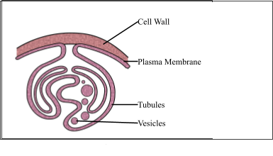
Mesosomes
When the plasma membrane folds, mesosomes are created. Respiratory enzymes are present in them. Lamellae, tubules, and vesicles make up their structure.
Mesosome Types
They fall into one of two categories:
1. Mesosome of the Septum
They are observed connected to the cell wall or chromosomes of dividing cells. It frequently plays a role in the development of cell walls, chromosome replication, and chromosomal distribution to daughter cells.
2. The Lateral Mesosome
They don’t appear to be connected to the nucleoside. It has enzymes for breathing.
Mesosome Functions
The following are mesosomes’ primary roles:
- Development of cell walls.
- Replication of DNA.
- DNA replication is transferred to the offspring cells.
- Increasing the plasma membrane’s surface area.
- Enzymatic and secretor activities.
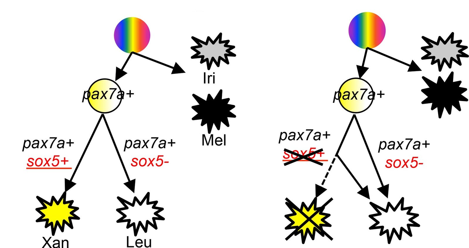
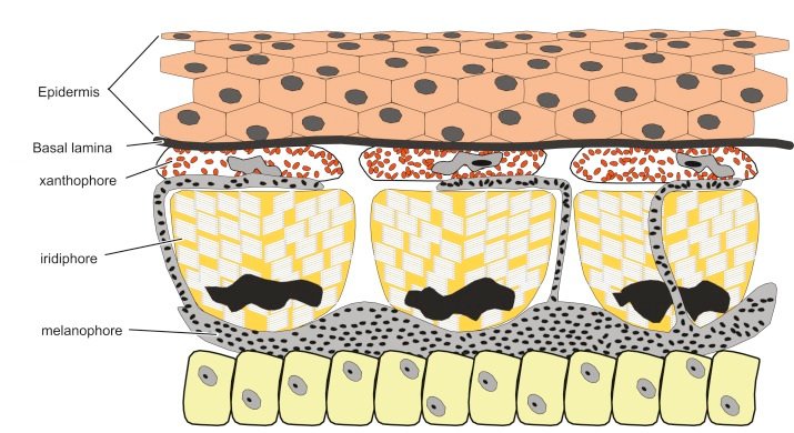
Chromatophores
These are the membranous extensions containing pigment molecules such as bacterial chlorophyll. These are abundantly found in cyan bacteria or blue green algae. Their main role is photosynthesis. They may be of different shapes like lamellar, tubular or vesicular in shape.
Classification
There are three different categories of chromatophores:
- Iridophores: These cells have reflective platelets, which alter how they are oriented to reflect various colors.
- Melanophores: The melanin that gives the skin its dark hues is found in these cells.
- Xanthophores: The arytenoids that give the skin its yellow and orange hues are found in these cells.
Both xanthophores and erythrophores
Depending on whether they contain a large amount of yellow pteridine pigments or mostly red/orange arytenoids, chromatophores are categorized as either erythrophores or xanthophores. However, when vesicles containing pteridine and carotenoids are present in the same cell, the total color depends on the ratio of red and yellow pigments. As a result, it’s not always easy to tell these chromatophores apart.
Applications & Functions
Animals’ skin contains chromospheres, which are pigment cells that change color. They are used for camouflage, temperature control, and communication. Chromospheres contain a pigment called melanin, which absorbs light. When the pigment cells are constricted, the concentrated melanin makes them seem dark. As the cells proliferate, the melanin is dispersed, giving them a light appearance.
- Chromatophores are frequently employed in applied research. For instance, the coordination and communication of chromatophores to create the continuous horizontal striped pattern observed in adult fish is studied in zebra fish larvae.
- In evolutionary developmental biology, this model system is thought to be useful for comprehending patterning.
- Additionally, chromatophore biology has been used to represent human illnesses or disorders such as melanoma and albinism.